In discussions surrounding breast cancer detection, the terms sensitivity and specificity hold paramount importance. A systematic review and meta-analysis conducted in 2019 revealed that ultrasound boasts an impressive sensitivity of 80.1% and specificity of 88.4%. These metrics serve as essential benchmarks; sensitivity informs us of a test’s capability to identify true positives, meaning it effectively finds individuals who actually have breast cancer. On the flip side, specificity tells us how adeptly a test can rule out those who do not have the disease, resulting in fewer false positives.
Understanding these definitions allows us to appreciate the strengths and limitations of ultrasound as a diagnostic tool. While it is not foolproof in identifying every instance of breast cancer, its high levels of sensitivity can be a game-changer for patients, particularly those presenting complex cases or difficult-to-detect anomalies.
When Is Ultrasound Recommended?
Breast ultrasound is not merely an adjunctive procedure; it plays a pivotal role in comprehensive breast cancer screening. According to Cancer Research UK, healthcare professionals recommend ultrasound as a first-line test under a variety of conditions. For instance, women with palpable lumps that mammograms have failed to detect often undergo ultrasounds for further examination. It is also invaluable in distinguishing between solid masses and cysts, thereby guiding treatment options. Additionally, individuals with dense breast tissue—where mammograms might fall short—can greatly benefit from ultrasound screenings.
The recommendation from healthcare providers to combine breast ultrasound with mammograms and physical exams signifies its importance in the early detection process. This combined approach not only enhances diagnostic accuracy but fosters a thorough investigative strategy to ensure no potential malignancies are overlooked.
The Ultrasound Experience: What Patients Should Expect
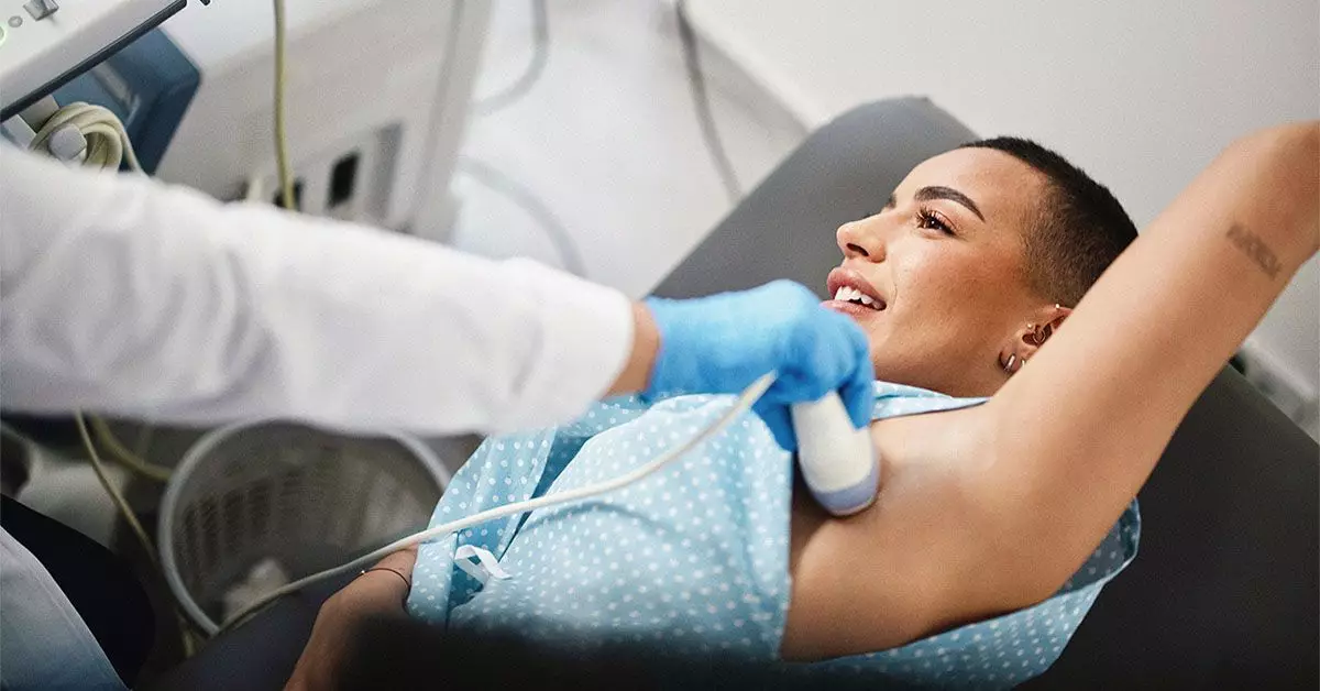
For anyone preparing to undergo a breast ultrasound, understanding the process can alleviate anxiety. During the assessment, patients typically remove clothing from their top half and have a lubricating gel applied to their breast. This gel is crucial; it optimizes sound wave transmission for clearer images and more accurate readings. The sonographer—the trained professional conducting the test—will deftly maneuver the ultrasound probe, capturing images not just of the breast, but also the surrounding lymph nodes in the armpit region.
This non-invasive process is quick and generally well-tolerated, making it an accessible form of screening for many women. The ultrasound examination further allows for real-time evaluations, giving providers immediate insights that may necessitate further intervention, such as a biopsy.
Shifting Guidelines for Breast Cancer Screening
The evolving guidelines around breast cancer screening highlight the dynamic nature of medical recommendations. The American Cancer Society endorses annual screenings starting at age 45 for individuals with average risk, while providing the option for those aged 40 to 44 to initiate annual mammograms if desired. This presents an opportunity for proactive monitoring, empowering women to take charge of their breast health.
Such guidelines emphasize the importance of vigilance in detecting the disease early—an endeavor made more effective with the incorporation of ultrasound technology. Women noticing any unusual changes in their breasts are strongly encouraged to consult healthcare professionals, reinforcing a culture of communication and awareness in breast cancer detection.
By embracing and understanding the pivotal role of ultrasound in breast cancer screening, we can foster a more informed community. The transparency provided by these discussions around sensitivity, specificity, and the ultrasound process could ultimately save lives, highlighting the importance of proactive health measures in the fight against breast cancer.

